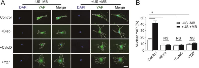
Figure 7.

Actin, myosin II, and RhoA/ROCK signaling are all required for rescuing YAP activity in hMSCs on soft substrates by ATC. (A&B) Representative fluorescence images (A) and bar plot (B) showing subcellular YAP localization in hMSCs seeded on a soft PMA (effective substrate rigidity Eeff = 1 kPa) under different drug treatments as indicated. hMSCs were coated with functionalized microbubbles (MBs) before ultrasound (US) stimulation (+US +MB). Negative control for hMSCs without MBs and US stimulation (−US −MB) was included for comparison. Cells were treated with pharmacological drugs for 1 hr before US application. Blebbistatin, or Bleb, 50 μM; Cytochalasin D, or CytoD, 40 μM; Y-27632, or Y27, 50 μM. Cells were stained for YAP 30 min after US stimulation. White dashed lines in micrographs highlight cell boundaries. Scale bar, 50 μm. Error bar, the mean ± s.e.m, with n ≥ 3. *, P < 0.05; NS, P > 0.05. US parameter: frequency 1 MHz, pulse repetition frequency 10 Hz, pulse duration 50 ms, treatment duration 5 min, and acoustic pressure 0.08 MPa.
