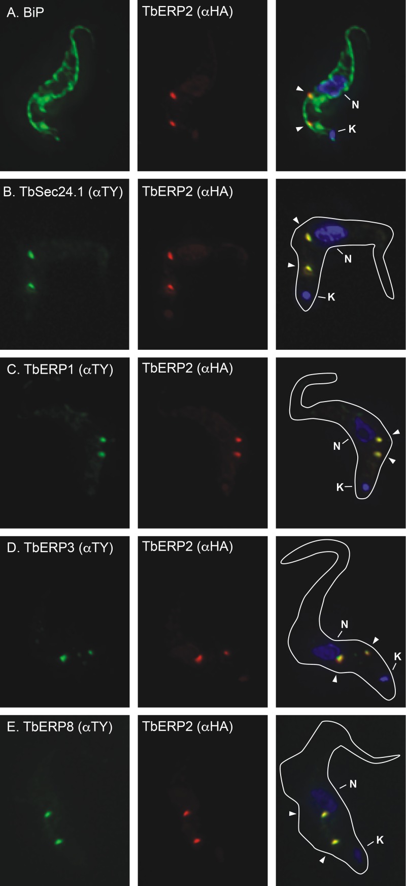
FIG 1 .
TbERP1, TbERP2, TbERP3, and TbERP8 colocalize at ER exit sites. The in situ TY tagging constructs for TbSec24.1 (B), TbERP1 (C), TbERP3 (D), and TbERP8 (E) were each transfected into the BSF HA-TbERP2 cell line (A), generating a panel of differentially tagged cell lines as indicated. The various cell lines were fixed, permeabilized, and stained with anti-BiP or anti-TY (left, as indicated) and anti-HA (middle). Cells were then stained with appropriate secondary antibodies (HA, red; BiP or TY, green) and DAPI to visualize nucleic acid (blue). Three-channel merged images and cell body outlines from corresponding DIC images are presented (right). All images are deconvolved sum-stack projections of representative G1-phase cells. Arrowheads, TbERP-positive foci; N, nucleus; K, kinetoplast.

