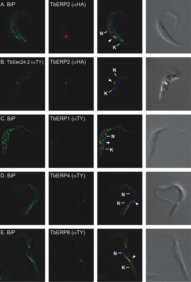
FIG 7 .
TbERP1, TbERP2, TbERP4, and TbERP8 localize to ERES in PCF cells. The in situ tagging constructs for TbERP2 (A), TbERP2 and TbSec24.2 (B), TbERP1 (C), TbERP4 (D), and TbERP8 (E) were transfected into the PCF cell line. Cells were fixed, permeabilized, and stained with anti-BIP, anti-TY, and/or anti-HA as indicated. Cells were then stained with appropriate fluorescently labeled secondary antibodies (BiP, green; HA, red; TY, green or red) and DAPI to visualize nucleic acid (blue). Three-channel merged images and corresponding DIC images are presented at right. All images are deconvolved sum-stack projections of representative G1-phase cells. Arrowheads, TbERP-positive foci; N, nucleus; K, kinetoplast.

