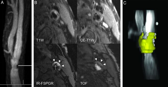
Fig 1.
A 75-year-old man with asymptomatic mild left carotid artery stenosis and complex carotid plaque. A, Maximum intensity projections from contrast-enhanced MRA show irregular narrowing of the distal left common carotid artery with stenosis of 33% diameter. The horizontal line indicates the level of transverse carotid plaque imaging. B, The chevrons demonstrate a large nonenhancing LR/NC region on CE-T1WI. Much of the LR/NC contains IPH seen as bright on IR-FSPGR and TOF. Last, a fibrous cap rupture with an ulcer penetrating into the hemorrhagic LR/NC is seen as depicted by the thin arrow. C, The large LR/NC is yellow, and calcifications are white on this volume-reformatted image generated from the multicontrast carotid plaque series. The total volume of LR/NC is 137 mL, and the percentage of the maximum area of LR/NC is 47%. Such large-volume LR/NC regions are frequently associated with other complex carotid plaque features such as IPH and/or fibrous cap rupture that correspond to American Heart Association type VI carotid plaque, which was found to occur significantly more often in men than in women.

