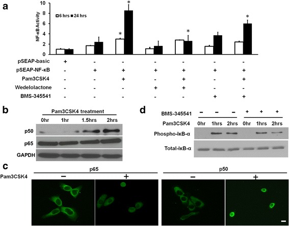
Fig. 1.

a NF-κB transcriptional activity assay. HCET cells were transfected with pSEAP-basic or pSEAP-NF-κB vector, treated with Pam3CSK4 (a TLR2 ligand) for 6 and 24 h with or without the presence of IκK inhibitors Wedelolactone and BMS-345541. Height of each bar represents mean of two independent experiments and is normalized to negative control (pSEAP-basic). The error bars represent standard error of the means. b HCE-T cells were treated with Pam3CSK4 for various time intervals and nuclear fraction of treated cells were immunoblotted with antibodies against p50 and p65. c HCE-T cells seeded in chamber slides were treated with Pam3CSK4 for 4 h and immunofluorescent staining was performed using primary antibodies against p65 and p50, and Alexa Fluor 488 conjugated secondary antibody (green). Representative images are shown. Scale bar = 50 μm. d. HCE-T cells were treated with Pam3CSK4 with or without IκK inhibitors for various time intervals and total cell lysate was immunoblotted with antibodies against phospho-IκB-α and total IκB-α. GAPDH was used as the endogenous loading control. *: P < 0.05
