
Figure 3. Glutamate pre- and post-synaptic marker expression is altered in T. gondii- infected mice at 8 wpi.
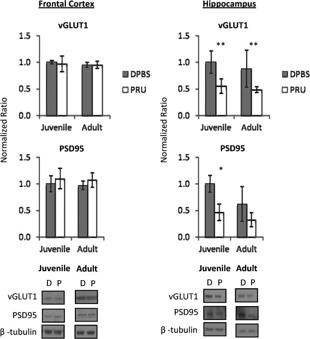
Frontal cortex pre-synaptic vesicular glutamate transporter 1 (vGLUT1) and post-synaptic density 95 (PSD95) expression were unchanged by infection. Hippocampal vGLUT1 was decreased in juvenile- and adult- infected mice. Linear regression analysis revealed no age × infection interaction. Hippocampal PSD95 was decreased in juvenile-infected mice and an age × infection interaction was determined (b=-0.261, t=-2.435, p=0.026). Data represent an average of 5 mice per group and 4 western blots per sample, with representative western blot images shown. DPBS (D) denotes mock-infected controls and PRU (P) denotes T. gondii infected mice. Bars represent 95% confidence interval and symbols represent statistical significance from Mann-Whitney U-test comparisons.
* denotes p≤ 0.05 compared to age-matched mock-infected (DPBS) group
** denotes p≤ 0.01 compared to age-matched mock-infected (DPBS) group