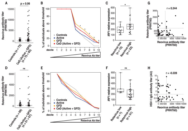
Fig. 5. Role of human reovirus infections in celiac disease pathogenesis.
(A and D) Boxplots showing levels of reovirus antibody titers (A) and rotavirus antibody titers (D) in control (n = 73) and CeD patients (n = 160). (B and E) Percentage of control patients (gray), active CeD patients (blue), CeD patients on a gluten-free diet (GFD) (orange), and active CeD and GFD patients combined (red) that have reovirus (B) or rotavirus (E) antibody titers above an increasingly higher cutoff (left to right). Cutoffs were determined by defining the deciles of the distribution of reovirus (B) or rotavirus (E) antibody titers observed in the patient samples analyzed. GFD and CeD (Active + GFD) patient groups are significantly overrepresented among individuals with reovirus titers above a PRNT60 = 156 (6th decile) and PRNT60 = 1597 (9th decile), respectively. (C and F) IRF1 expression in small intestinal biopsies of GFD patients (n = 38) was analyzed by means of RT-PCR. To avoid any confounding factors associated with increased inflammation, analysis of IRF1 expression was performed in CeD patients on a GFD who, unlike active CeD patients, display normal levels of IFN-γ. Relative expression level of IRF1 in GFD patients with low (left) and high (right) levels of reovirus (C) or rotavirus (F) antibody titers is shown. (C) Reovirus low and reovirus high were respectively defined as individuals with antibody titers below and above the median PRNT60 = 47 [(B), 5th decile]. (F) Rotavirus low and rotavirus high were respectively defined as individuals with antibody titers below and above the median PRNT60 = 53 [(E), 5th decile]. (G and H) Reovirus and rotavirus antibody titers in serum of patients were determined by means of plaque-reduction neutralization assay. Levels of HSV-1 antibody titers were determined by means of ELISA. Correlations between the levels of reovirus antibody titers and rotavirus antibody titers (G) or HSV-1 antibody titers (H) in GFD patients are shown. r, Pearson correlation. (A) to (F), *P < 0.05; Mann-Whitney U test.

