Figure 2.
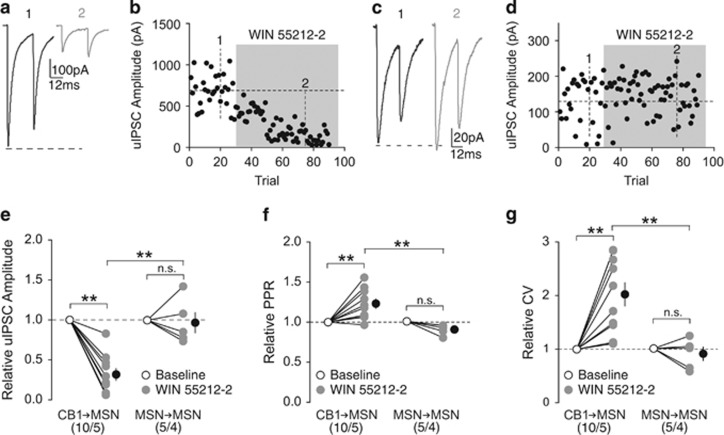
CB1 signaling preferentially suppresses inhibitory input from CB1+ FSIs. (a, b) uIPSC traces (a) and time course of uIPSC amplitude (b) from an example of functionally connected CB1-to-MSN pair before (1) and during (2) perfusion of WIN 55212-2 (5 μM). (c, d) uIPSC traces (c) and time course of uIPSC amplitude (d) from an example of functionally connected MSN-to-MSN pair before (1) and during (2) perfusion of WIN 55212-2 (5 μM). (e) Summary showing perfusion of WIN 55212-2 decreased the amplitude of uIPSCs at CB1-to-MSN synapses, whereas uIPSCs at MSN-to-MSN synapses were insensitive to WIN 55212-2. (f) Summary showing that perfusion of WIN 55212-2 increased the PPR of uIPSCs at CB1-to-MSN synapses, whereas uIPSCs at MSN-to-MSN synapses were unaffected. (g) Summary showing that perfusion of WIN 55212-2 increased the CV of uIPSC responses at CB1-to-MSN synapses, whereas uIPSCs at MSN-to-MSN synapses were unaffected. n/m represents number of cells/number of animals. **p<0.01. Error bars represent SEM.

