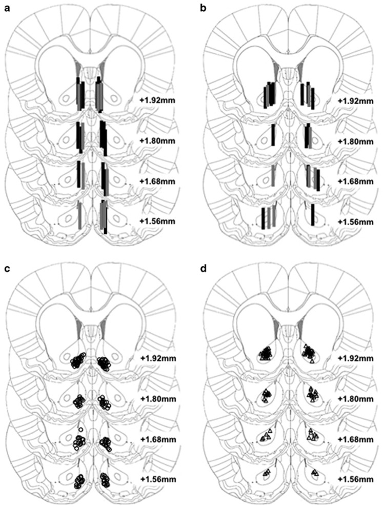
Figure 1.
Cannula placements for all experiments. Approximate locations of active region of microdialysis probe targeting the NAc shell (a) for Experiment 1A (n=18; black rectangles) and Experiment 1B (n=9; gray rectangles), or the NAc core (b) for Experiment 2A (n=14; black rectangles) and Experiment 2B (n=9; gray rectangles). Approximate anatomical position for microinjector tips targeting the NAc shell (c) for Experiment 3 (n=66; open circles) or NAc core (d) for Experiment 4 (n=46; open triangles). Images modified from the brain atlas of Paxinos and Watson (2005) of Figures 17–20 (+1.56 to +1.92 mm anterior to Bregma).

