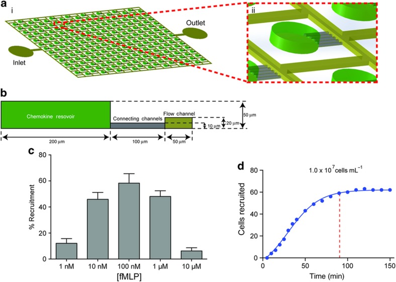
Figure 1.
A microscale device for studying neutrophil chemotaxis. (a) Schematic of the microfluidic device used for neutrophil chemotaxis assays. (ii) Magnified view of the reservoir representing one of the chemotaxis cells. (b) Geometric representation of chemotaxis cell. (c) Variation of neutrophil motility for different concentrations of formyl-methionyl leucyl-phenylalanine (fMLP). Maximum recruitment of ~60% of cells was observed for [fMLP]=100 nM. Significant number of neutrophils were recruited for starting concentration in the reservoirs between 10 and 1000 nM. (N=3. Error bars=mean±standard error of the mean). (d) Recruitment of neutrophils in response to fMLP. Recruitment peaked at ~90 min.

