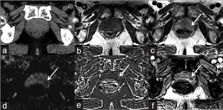
Figure 1.

An 81-year-old male with prostate cancer in the left peripheral zone. Prostate cancer and hemorrhage are not displayed on the CT image (a), isointensity on T1WI (b), iso- to hypointensity on T2WI (c) and ADC map (d). Arrows indicate the areas of prostate cancer. Hypointensity on susceptibility-filtered phase images (e), and susceptibility-weighted image (f) (arrows), prove the micro-abnormality of prostate cancer.
