
Figure 2.
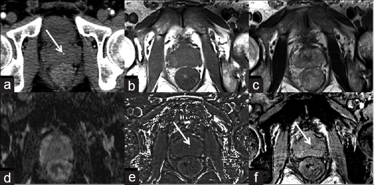
A 58-year-old male with calcification in the central zone of prostate. Prostatic calcification is seen as a dot-like high-density spot on the CT image (a) (arrow), but is hard to be identified on conventional T1WI (b), T2WI (c), and ADC map (d). Hyperintensity on susceptibility-filtered phase images (e) and hypointensity on susceptibility-weighted image (f) (arrows) indicate calcification.