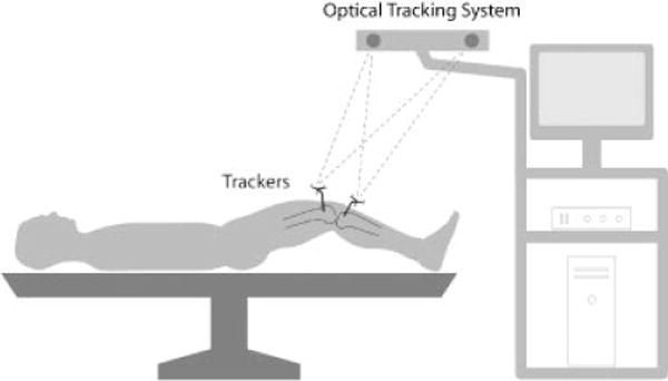
Figure 1.

Intraoperative experimental setup. An optical tracking system recorded knee motions using the positions and orientations of trackers affixed to the femur and tibia. At four surgical stages, knee motions were recorded as the surgeon moved the knee through its range of flexion and extension. (see methods for details)
