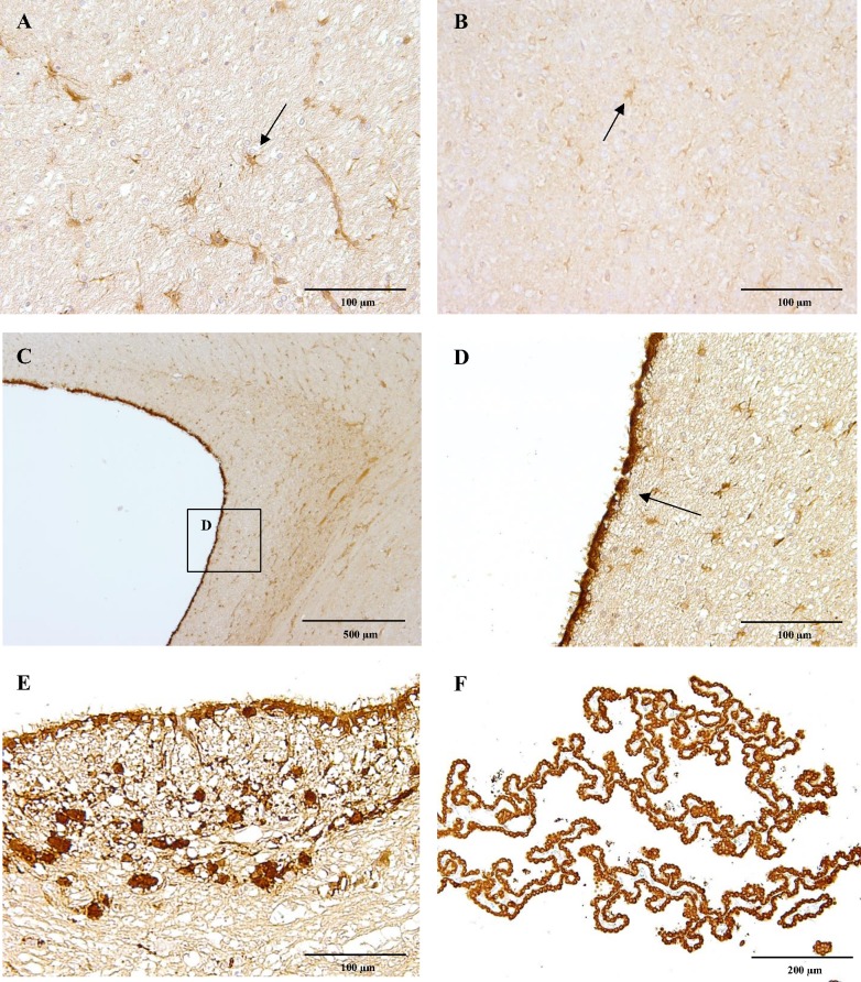
Fig 1. CB1 immunoreactivity.
Astrocytes (arrow) of the cerebral white matter of a six-month-old Beagle dog showing strong CB1 receptor immunoreactivity (A) comparing to astrocytes of a four-week-old dog, which are only slightly positive (B). The ependymal cells (arrow) of a six-month-old dog lining the lateral ventricle strongly express CB1 receptor (C, D). Similarly, ependymal cells lining the fourth ventricle and scattered neuroglial cells (E) are CB1 receptor positive, as well as cells of the choroid plexus (F). IHC was performed using the avidin-biotin-peroxidase complex (ABC) method.

