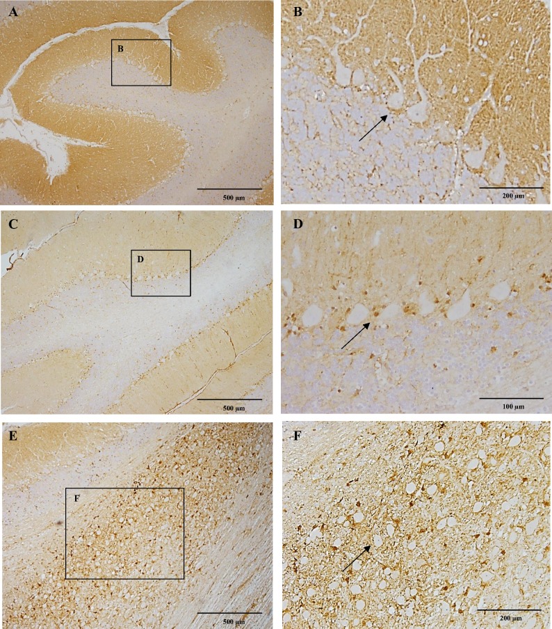
Fig 4. CB1 immunoreactivity of the cerebellum and choclear nuclei.
In figure A notice strong CB1 immunoreactivity within the molecular layer of the cerebellar cortex in a six-month-old Beagle dog. Figure B depicting in detail immunonegative Purkinje cells surrounded by strong immunorreactive fibers particularly in the basal portion (arrow). In the ten-year-old dog, there is a slight immunoreactivity in the molecular layer of the cerebellar cortex (C). Purkinje cells surrounded by a dot-like immunoreactivity appear devoid of immunoreactivity in the ten-year-old dog (D; arrow). The cochlear nucleus in a six-month-old dog showing strong CB1 immunoreactivity (E). In figure F detail of the cochlear nucleus with strong CB1 immunoreactivity surrounding the unstained neuronal bodies (arrow). IHC was performed using the avidin-biotin-peroxidase complex (ABC) method.

