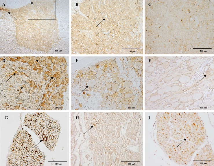
Fig 5. CB1 immunoreactivity in the spinal cord, dorsal root ganglia and peripheral nerve.
In figure A, strong CB1 immunoreactivity is shown in the grey matter of the cervical spinal cord of a six-month-old dog and the cytoplasm of ependymal cells lining the central canal (A; arrow). Within the dorsal horn, CB1 immunoreactivity appears surrounding unstained neuronal bodies (B; arrow). In the cervical spinal cord of a ten-year-old dog notice slight immunoreactivity of the grey matter (C). Figure D showing the cervical dorsal root ganglia of a six-month-old dog with slight immunoreactivity of large neurons and strong CB1 immunoreactivity of small dark neurons (arrows) and satellite cells (arrowheads). The thoracic dorsal root ganglia of a ten-year-old dog with moderate CB1 immunoreactivity of small dark neurons and satellite cells, large neurons show slight immunoreactivity (E; arrow). The cervical dorsal root ganglia of a four-week-old dog depicting scattered large and small neurons and satellite cells with slight CB1 immunoreactivity (F; arrow). In figure G the cervical spinal nerve of a six-month-old dog shows strong CB1 expression in Schwann cells ensheating axons (arrow). Few Schwann cells show moderate CB1 immunoreactivity (arrow) in a thoracic spinal nerve of a ten-year-old dog (H). The cervical spinal nerve in the four-week-old dog shows moderate CB1 immunoreactivity of scattered Schwann cells (I; arrow). IHC was performed using the avidin-biotin-peroxidase complex (ABC) method.

