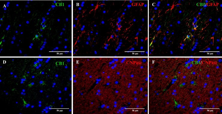
Fig 6. Double immunofluorescence staining of the cerebral white matter of a six-month-old Beagle dog.
Double immunofluorescence staining of CB1 (green, A) with GFAP (red, B) reveals co-localization in about 20% astrocytes (C). CNPase expression (red, E) and CB1 (green, D) do not co-localize, suggesting a lack of expression of CB1 receptors by mature oligodendrocytes (F). Nuclear staining (blue) with bisbenzimide.

