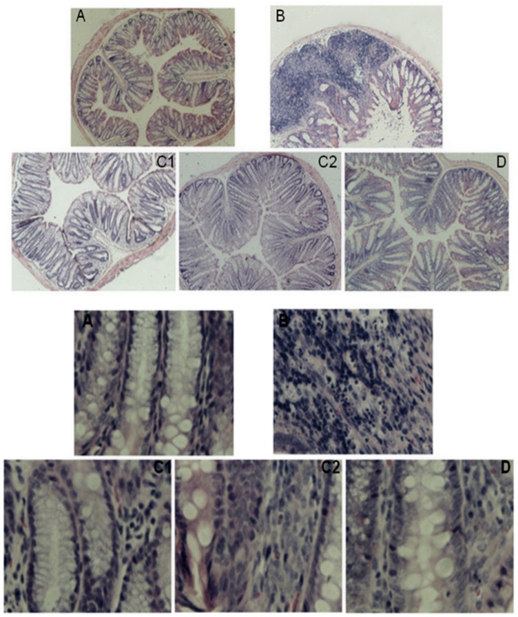
Fig 2. Images of intestinal histopathological sections from mice in the various groups: H&E staining with magnification at 40x and 400x.
One-centimeter sections of normal and obviously pathologically altered intestine were removed from mice in each group, and paraffin sections were prepared using routine procedures, including paraffin embedding, sectioning, H&E staining and mounting with neutral balsam. Histopathological changes were observed with an inverted microscope. A: normal control group; B:OXZ-induced UC model group; C1: CGMP group 1 (50 mg/kg bw•d); C2: CGMP group 2 (200 mg/kg bw•d); and D: SASP-treated group. The upper image contains “A, B, C1,C2 and D” were based on images of H&E-stained sections at 40x magnification and The lower image contains bold “A, B, C1,C2 and D” were based on images of H&E-stained sections at 400x magnification.

