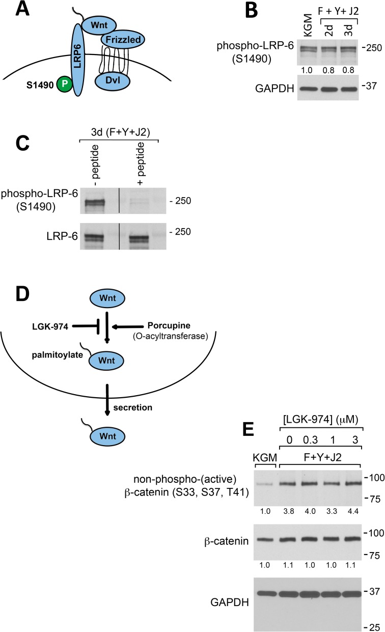
Fig 2. Wnt signaling does not activate β-catenin in CR HECs.
(A) Diagram showing Wnt-dependent phosphorylation of LRP6 at S1490. (B) Western blot analysis of LRP6 phosphorylation at S1490 in whole-cell lysates prepared from HECs in KGM (KGM) after 3 d, or from 2–3 d CR cultures (F+Y+J2). All cultures included 0.15% DMSO. (C) Western blot of phospho-LRP6 (S1490) and total LRP6 from 3 d HEC CR cultures with and without pre-incubation of the primary antibody with a blocking peptide recognizing the S1490 phosphorylation site (see Materials and methods). (D) Schematic drawing showing LGK-974 inhibition of Wnt palmitoylation by Porcupine, which is obligatory for Wnt secretion. (E) Western blot analysis of β-catenin activation in whole-cell lysates prepared from HECs in 3 d KGM cultures (KGM), or from 3 d CR cultures in the presence of 0–3 μM LGK-974. All cultures included 0.15% DMSO. In (B), (C) and (E), lanes contain equal amounts of protein. Molecular mass markers (in kDa) are shown on the right.

