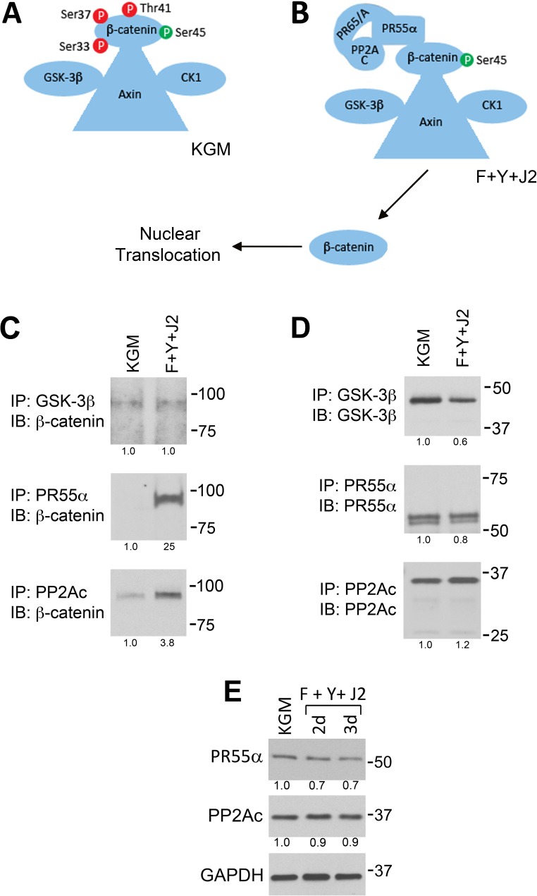
Fig 5. Increased association of PP2A regulatory and catalytic subunits with β-catenin in CR HECs.
(A) Graphic illustration of the β-catenin complex in KGM. (B) Illustration of the β-catenin complex in CR cells (F+Y+J2). In (A) and (B), the β-catenin priming phosphorylation site at S45 is green, while destabilizing phosphorylation sites are red. (C) GSK-3β, PR55α and PP2Ac were immunoprecipitated (from equal amounts of protein) from HECs in KGM cultures (KGM), or from CR cultures (F+Y+J2) after 3 d. The immunoprecipitates (IP) were analyzed on immunoblots (IB) labeled for β-catenin to measure association between the proteins. (D) Control experiments (IP and IB the same protein) verified that equivalent amounts of each protein were immunoprecipitated in (C). (E) Total levels of PR55α and PP2Ac in 3 d KGM cultures (KGM) and in HECs conditionally reprogrammed for 2–3 d (F+Y+J2). Lanes contain equal amounts of protein. In (C), (D) and (E) molecular mass markers (in kDa) are shown on the right.

