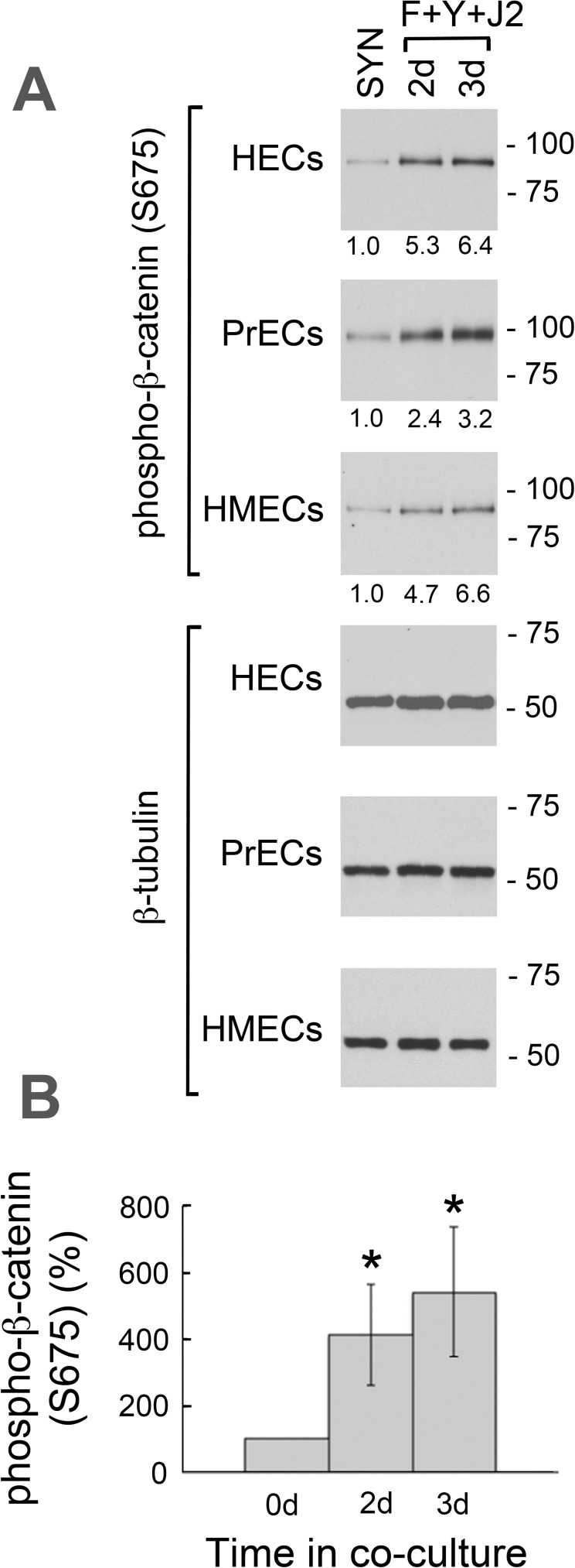
Fig 8. β-catenin phosphorylation at S675 in synthetic media and CR cultures.
(A) Whole-cell lysates were prepared from 3 d cultures of HECs, PrECs and HMECs in synthetic media (SYN: KGM for HECs and PrECs; MEGM for HMECs), or from 2–3 d CR cultures (F+Y+J2). Western blots were labeled for phospho-β-catenin (S675) and normalized relative to identical immunoblots labeled for β-tubulin. Lanes contain equal amounts of protein. Molecular mass markers (in kDa) are shown on the right. (B) Mean phospho-β-catenin (S675) levels in the 3 cell lines. Error bars indicate standard deviation (S.D.) from the mean. (*) P-value < .00001 relative to 0 d.

