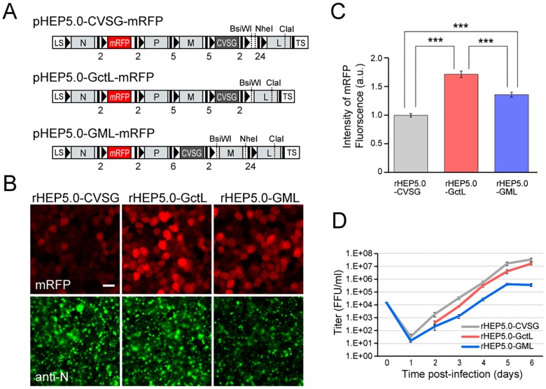
Fig 1. Characteristics of RV vectors in cultured cells.
A: Genome organization of recombinant RV vectors. The transcription start and stop/polyadenylation signals are respectively indicated by black bars and black arrowheads in the schematic diagram, and the number of nucleotides of IGR are shown below the diagram. LS, leader sequence; TS, trailer sequence. B: Photomicrographs of RV-infected NA cells at 2 dpi. The three RV vectors expressed the transgene mRFP (red), which was inserted between the N and P genes. Infection of the viral vector can be confirmed by immunofluorescence of the N protein (green). Scale bar = 20 μm. C: Fluorescence intensities of mRFP in infected cells at 2 dpi [mean ± standard errors, numbers of analyzed cells: 189 (rHEP5.0-CVSG), 382 (rHEP5.0-GctL), 276 (rHEP5.0-GML), *** p < 0.001]. D: Viral titer growth curves of RV vectors (mean ± standard errors, N = 6). No viruses were detected in rHEP-5.0-GctL-infected cells at 1 dpi.

