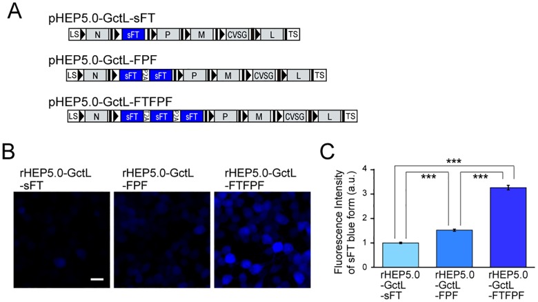
Fig 3. Characteristics of sFT-expressing RV vectors in vitro.
A: Genome organization of recombinant RV vectors. B-C: Fluorescence photomicrographs of NA cells infected with sFT-expressing RV vectors (B) and fluorescence intensity of sFT in infected cells [mean ± standard errors, numbers of analyzed cells: 1199 (rHEP5.0-GctL-sFT), 924 (rHEP5.0-GctL-FPF), and 646 (rHEP5.0-GctL-FTFPF), *** p < 0.001] at 2 dpi (C). Scale bar = 20 μm.

