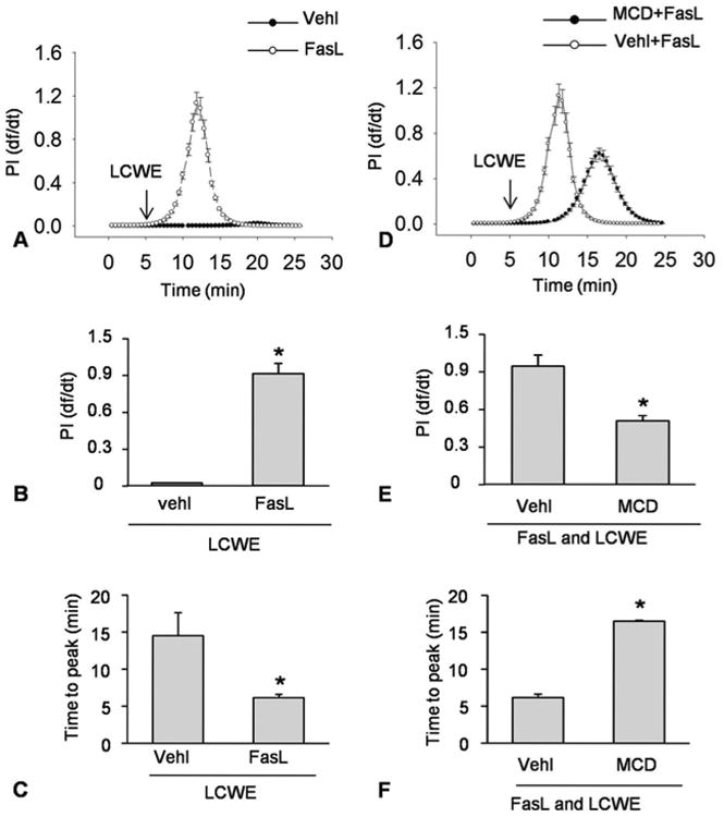
Figure 3.

Enhanced MR clustering blocked membrane resealing during LCWE-induced injury. MVECs were stimulated without (control: ctrl) or with LCWE (15 μg/ml) in the absence and presence of MR clustering inducer (FasL) and MR clustering inhibitor (MCD). A and B) Representative curves depicting the relationship of FasL enhanced LCWE-induced the degree of plasma membrane injury to the flow velocity of PI by change in relative fluorescence with time (df/dt) of PI fluorescence. C and D) Representative histograms showing the maximal df/dt (N=6). E and F) Representative histograms presenting the time to reach maximal df/dt (N=6). *P < 0.0.5 vs. LCWE or FasL + LCWE.
