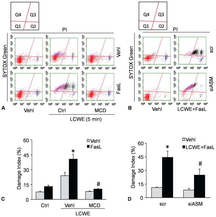
Figure 4.

LCWE-induced plasma membrane injury and repair in MVECs analyzed by flow cytometry. MVECs were stimulated without (control: ctrl) or with LCWE (15 μg/ml, 4 h) in the absence and presence of MR clustering inducer (FasL) and MR clustering inhibitor (MCD). A and B) Representative dot plots of sorted cells by flow cytometry, where MVECs were first stained by the SYTOX dye and the by PI to determine cell viability. Summarized data in panels C and D showing the cell number of Q4 area. *P < 0.0.5 vs. vehicle control; #P < 0.0.5 vs. vehicle + FasL + LCWE.
