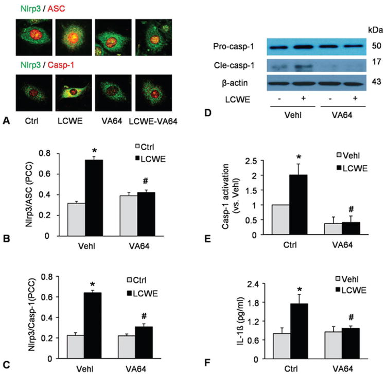
Figure 5.

Role of decreased cell plasma membrane resealing in activation of Nlrp3 inflammasome by LCWE. MVECs were stimulated without (control: ctrl) or with LCWE (15 μg/ml, 8 h) in the absence and presence of artificial cell membrane resealing reagent (VA64). A) Confocal microscopic images of MVECs stained with Alexa488-conjugated ant-Nlrp3 and Alexa555-conjugated anti-ASC or anti-caspase-1 antibodies. The co-localization is shown by yellow and Nlrp3 signal is green with ASC signal in red. or Nlrp3 in green vs.) caspase-1 in red. B and C) Summarized data showing the co-localization coefficiency of Nlrp3 with ASC or with caspase-1 (N=4). D-E) Western blot analysis of cleaved caspase-1 and pro-caspase-1 in MVECs (N=3). F) IL-1beta production in MVECs by ELISA (N=5). *P < 0.0.5 vs. vehicle control; #P < 0.0.5 vs. vehicle + LCWE.
