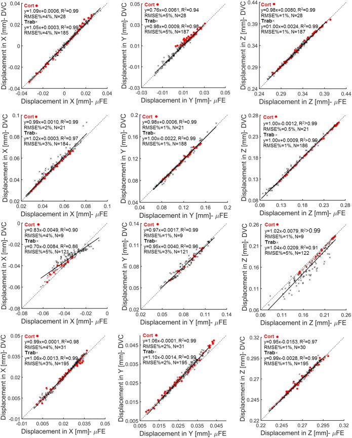
Fig 3. Regression analysis of microFE models predictions of local displacements per specimen and bone type.
MicroFE models predictions and DVC measurements computed along the transverse (X, Y) and axial (Z) directions for each specimen within cortical (red circles) and trabecular (black crosses) bone regions.

