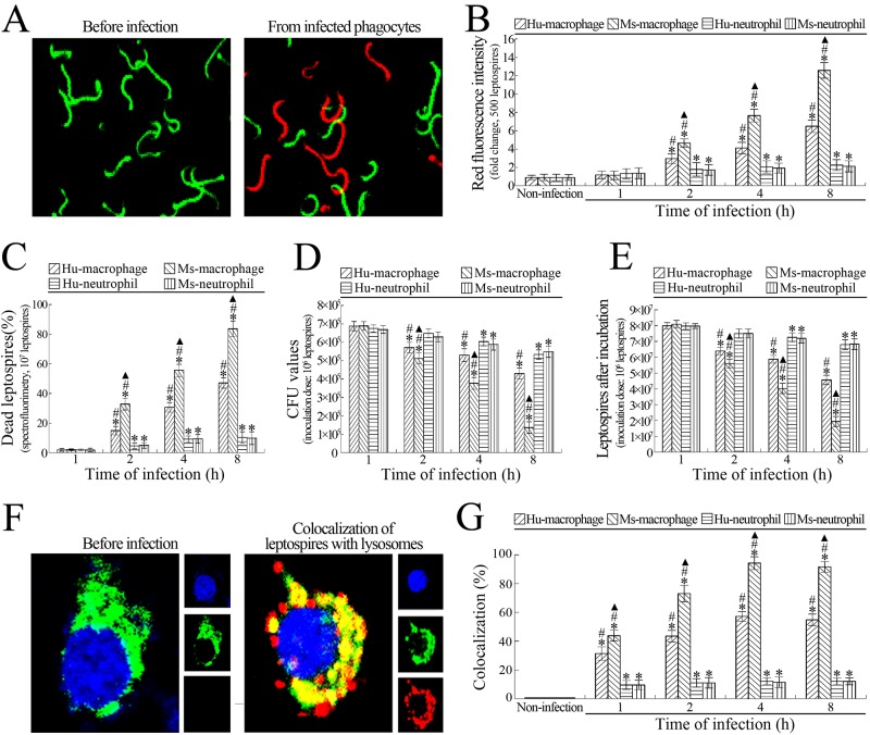
Fig 2. Macrophages have a higher ability of killing leptospires than neutrophils.
(A) Living and dead leptospires under a confocal microscope. The green leptospires were living while the red leptospires were dead. (B) Ability of macrophages and neutrophils to kill intracellular leptospires during infection with L. interrogans, determined by confocal microscopy for the indicated infection times. Bars show the means ± SD of three independent experiments. Five hundred leptospires in each experiment were analyzed to quantify the values of red fluorescence intensity. The means of red fluorescence intensities from the leptosires without infection were set as 1.0. *: p<0.05 vs the red fluorescence intensities of leptospires without infection. #: p<0.05 vs the red fluorescence intensities reflecting the dead leptospires from the infected Hu- or Ms-neutrophils. ▲: p<0.05 vs the red fluorescence intensities reflecting the dead leptospires from the infected Hu-macrophages. (C) Percentages of dead leptospires from macrophages and neutrophils during infection with L. interrogans, determined by spectrofluorimetry for the indicated infection times. Bars show the means ± SD of three independent experiments. 107 leptospires in each experiment were used to determine the dead leptospiral percentages. *: p<0.05 vs the dead percentages of the leptospires without infection. #: p<0.05 vs the dead leptospiral percentages from the infected Hu- or Ms-neutrophils. ▲: p<0.05 vs the dead leptospiral percentages from the infected Hu-macrophages. (D) Fewer leptospiral colonies from L. interrogans-infected macrophages than neutrophils, assessed by CFU enumeration for the indicated infection times. Bars show the means ± SD of three independent experiments. 106 leptospires from each of the infected cells were serially diluted and then inoculated onto EMJH-agar plates for a three-week incubation at 28°C for CFU enumeration. *: p<0.05 vs the CFU values of the leptospires without infection. #: p<0.05 vs the CFU values of the leptospires from the infected Hu- or Ms-neutrophils. ▲: p<0.05 vs the CFU values of the leptospires from the infected Hu-macrophages. (E) Attenuated growth ability of leptospires from L. interrogans-infected macrophages than neutrophils, assessed by leptospiral enumeration after incubation. Bars show the means ± SD of three independent experiments. 106 leptospires from each of the infected cells were inoculated in EMJH liquid medium for one-week incubation at 28°C for leptospiral enumeration. *: p<0.05 vs the growth ability of the leptospires without infection. #: p<0.05 vs the growth ability of the leptospires from the infected Hu- or Ms-neutrophils. ▲: p<0.05 vs the growth ability of the leptospires from the infected Hu-macrophages. (F) Co-localization of intracellular leptospires with lysosomes under a confocal microscope. Three fluorescence images were merged in the left panels and separate fluorescence channels in the right panel. The blue plaques indicate the nucleus. The green plaques around the nucleus indicate the lysosomes. The red spots around the nucleus indicate the intracellular leptospires. The yellow spots or plaques indicate the co-localization of intracellular leptospires with lysosomes. (G) Co-localization of intracellular leptospires with lysosomes in L. interrogans-infected macrophages and neutrophils, determined by confocal microscopy for the indicated infection times. Bars show the means ± SD of three independent experiments. One hundred cells in each experiment were analyzed to quantify the intensities of yellow fluorescence. The means of yellow fluorescence intensities from the cells without infection were set as 1.0. *: p<0.05 vs the yellow fluorescence intensities of the leptospires without infection. #: p<0.05 vs the yellow fluorescence intensity reflecting the intracellular leptospire-lysosome co-localization in the infected Hu- or Ms-neutrophils. ▲: p<0.05 vs the yellow fluorescence intensity reflecting the intracellular leptospire-lysosome co-localization in the infected Hu-macrophages.

