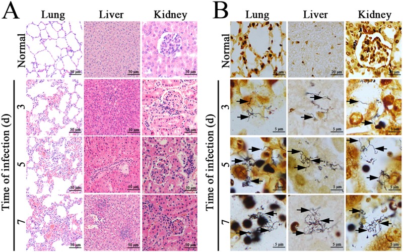
Fig 5. Histopathological changes and leptospires in tissues of L. interrogans-infected mice.
(A) Histopathological changes in lung, liver and kidney tissues from L. interrogans-infected C3H/HeJ mice, examined by microscopy after HE staining. All the tissues had infiltration of inflammatory cells. The lung, liver and kidney tissues presented serious hemorrhage, extensive hepatocellular necrosis and serious congestion, respectively. (B) Visible leptospires in lung, liver and kidney tissues from L. interrogans-infected C3H/HeJ mice, examined by microscopy after silver staining. The arrows indicate leptospires in the three types of tissues from the infected animals.

