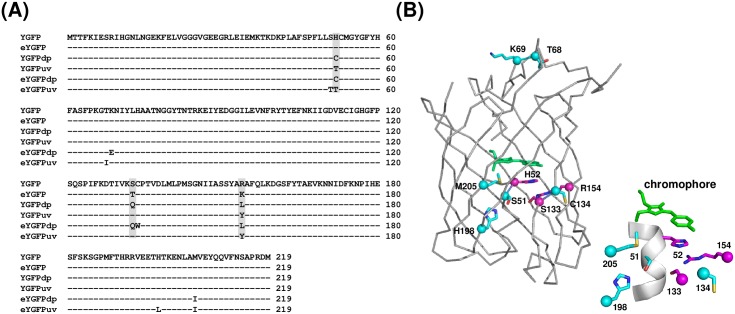
Fig 1. Amino acid sequence alignment of YGFP and its variants identified in this study.
(A) Shaded residues were replaced with random amino acids for the 1st library screening. (B) Overall structure of CpYGFP (Accession No. AB185173 and Protein Data Bank code 2DD7). The structure model was drawn using PYMOL software (DeLano Scientific; http://www.pymol.org). Amino acid mutations of eYGFPdp and eYGFPuv, both obtained from DNA shuffling, are shown as cyan spheres with side chains. The chromophore composed of a tripeptide, Gly-Tyr-Gly, in the center is shown in the stick model (green). Magenta spheres indicate the amino acids at positions 52, 133, and 154, which are described in (A). The sequences of these YGFP variants in this figure are available at the DDBJ/EMBL/GenBank nucleotide sequence databases with the accession numbers LC217529 (eYGFP), LC17530 (YGFPdp), LC217531 (YGFPuv), LC217532 (eYGFPdp) and LC217533 (eYGFPuv), respectively.

