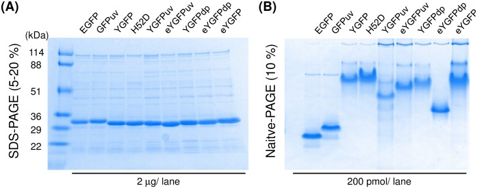
Fig 3. SDS-PAGE and native PAGE analysis of YGFP and its variants in comparison to EGFP and GFPuv.
(A) N-terminal His-tagged proteins were separated by SDS-PAGE in a 5–20% polyacrylamide gel under reducing conditions. The gel was stained with Coomassie blue. (B) Oligomeric states of YGFP derivatives or commercial GFP proteins were visualized after 10% non-denaturing gel electrophoresis. Data are representative of two independent experiments.

