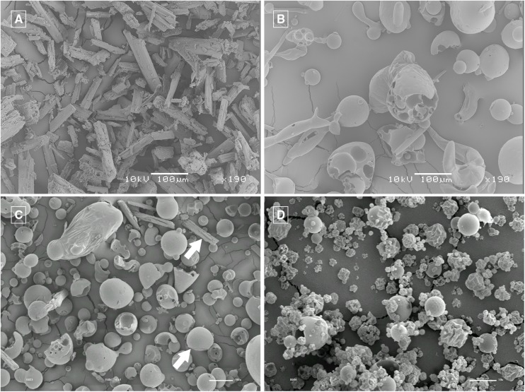
Fig 3. SEM micrographs.
Gallic acid (GA) 190X magnification (A), HPβCD 190X magnification (B), physical mixture of GA and HPβCD 150X magnification (C) and GA/HPβCD spray-dried microparticles, 2,000X magnification (D). In Fig C the upper arrow points out to the GA morphology and the lower arrow points out to the HPβCD one.

