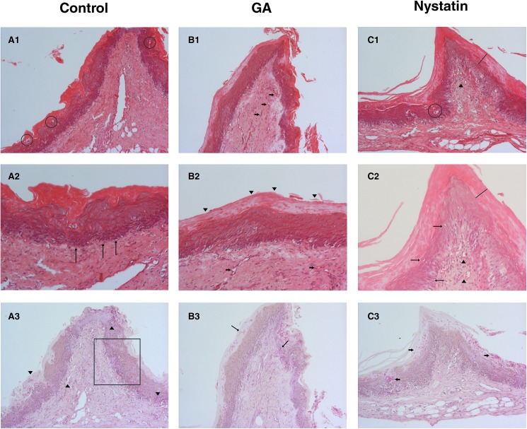
Fig 5. Antifungal activity on oral candidiasis in rats.
Control Group (A), GA/HPβCD spray-dried microparticles group (B) and Nystatin group (C). (A1) Histologic sections stained by Hematoxylin and eosin (HE) method, 100X magnification, exhibiting hyperkeratosis, and micro-abscesses formation (circle) in the epithelium that is also acanthotic. The fibrous connective tissue has wide vessels surrounded by a moderate inflammatory infiltrate. (A2) PAS staining, 100X magnification, where C. albicans yeast and mostly hyphae invade the epithelium reaching the connective tissue (arrowheads). (A3) HE staining, 200X magnification. Inflammatory epithelial alterations as hyperkeratosis, epithelial stratification loss (square), and basal layer disorganization (arrows) are evident. (B1) Histologic sections stained by hematoxylin and eosin (HE) method, 100X magnification, exhibiting hyperkeratosis, and acanthosis of the epithelium that covers a hyper-vascularized (arrows) connective tissue. (B2) HE staining, 200X magnification. Reactive hyperkeratosis of the epithelium (arrowheads) was the only remarkable inflammatory induced alteration for the GA/HPβCD group. Some congest vessels can be seen in the connective tissue (arrows). (B3) PAS staining, 100X magnification, where C. albicans yeast and mostly hyphae invade the epithelium, some of them reaching the connective tissue (arrows). (C1) Histologic sections stained by hematoxylin and eosin (HE) method, 100X magnification, hyperkeratosis (line), micro-abscess formation (circle), and acanthosis of the epithelium that covers a hyper-vascularized (arrowheads) connective tissue can be seen. (C2) PAS staining, 100X magnification where large groups of Candida albicans yeast and hyphae can be noted. Though most of them are limited to the keratin layer, some invade the epithelium (arrows). (C3) HE staining, 200X magnification, where epithelial hyperkeratosis (line) is evident. We can see poor inflammatory alteration (arrows), as hydropic degeneration and duplication of the basal layer. Some congest vessels can be seen in the connective tissue (arrowheads) surrounded by moderate inflammatory infiltrate.

