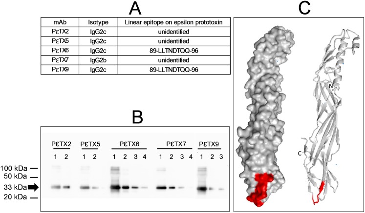
Fig 1. Characterization of mAbs.
A–Linear epitopes recognized by the different mAbs were identified as described in Methods. Antibodies for which no linear epitope was identified are categorized as “unidentified”, indicating that they probably bind a conformational epitope of epsilon prototoxin. B–Different quantities of epsilon prototoxin (lane 1: 1 μg, lane 2: 100 ng, lane 3: 10 ng, lane 4: 1 ng) were detected by western blotting with each of the different purified mAbs produced. The arrow indicates the prototoxin band (33 kDa). C–Localization of the linear epitope of mAbs PεTX6 and PεTX9 on the 3D-structure of epsilon prototoxin (surface (left) and ribbon (right) diagrams of the protein with accession code 1UYJ [33]).

