
Figure 5. Pathologic correlates of nonischemic scar.
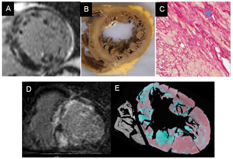
Panels A (pre-transplant CMR-LGE), B (post-transplant gross macroscopic cross-section) and C (post-transplant microscopic cross-section with fibrotic bundles, blue arrow) showing that the LGE with a midwall, near-circumferential pattern mirrors the distribution of pathologic replacement fibrosis. Panel D (pre-transplant CMR-LGE) shows diffuse LGE corresponding to regions of fibrosis confirmed by Masson’s trichrome staining (in green) on post-transplant histopathology (Panel E). E Reprinted from Iles et al.41 and Halliday et al.42 with permission of the publishers.