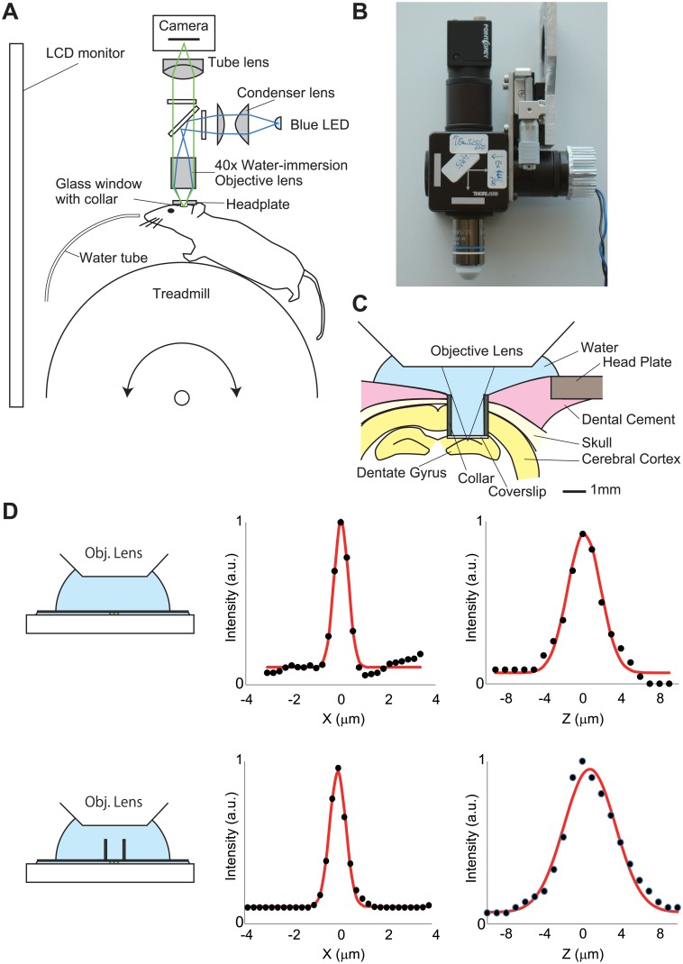
Fig 1. Wide-field imaging system for an awake head-restrained mouse.
(A) Simplified schematic of the endoscope imaging system. The blue line indicates the illumination pathway and the green line indicates the light collection pathway. Illumination light from a blue LED was collected with a condenser lens, passes through an excitation filter, reflects off a dichroic mirror, and irradiated through an objective lens. The fluorescence image was focused on a CMOS camera. (B) Image of the endoscope system. (C) Schematic of experimental setup showing the chronic window implant above the dentate gyrus. (D) Point spread function of the imaging setup with (lower) and without (upper) the cannula. Lateral (center) and axial (right) intensity line profiles across a 0.5 μm fluorescent bead (black dots) were shown. The read lines indicate Gaussian fits.

