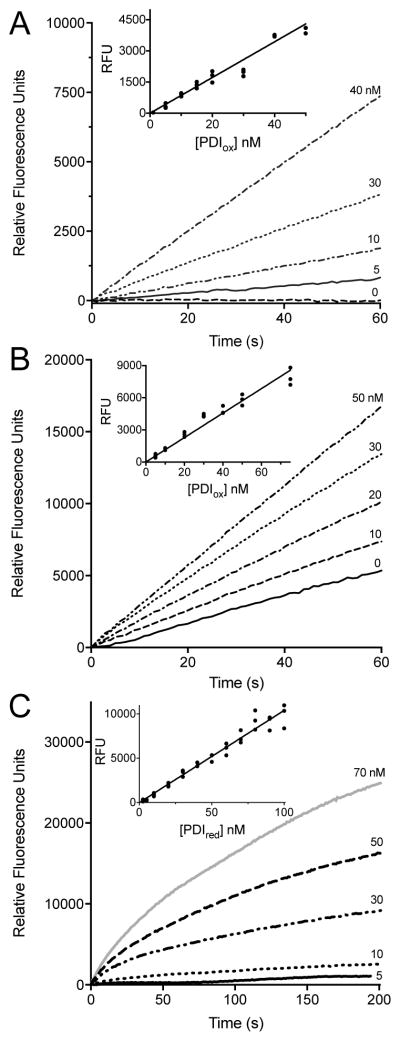
Fig. 4.
Multiple turnover and reoxidation PDI assays. Multiple turnover assays in buffer with (A) 5 μM DTT or (B) 5 mM GSH using increasing concentrations of oxidized PDI. The inset demonstrates the linearity of absorbance readings taken at 30 s as a function PDIox concentration, corrected for background thiol reduction of BD-SS. Assays were performed in triplicate, see Materials and Methods. (C) Reoxidation assays in buffer performed in triplicate using increasing concentrations of PDIred added to a final concentration of 714 nM BD-SS. Inset depicts absorbance readings at 30 s plotted against PDIred concentration.

