
Figure 1.
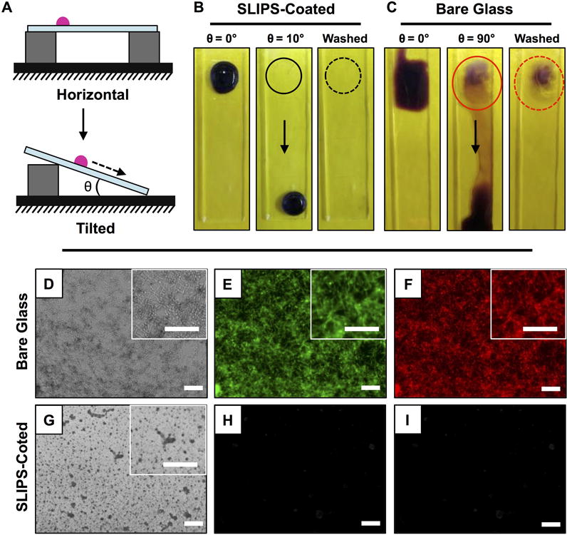
(A) Schematic illustration showing a side-view depiction of the beading and sliding of an aqueous droplet on horizontally placed (top) and tilted (bottom) ‘slippery’ liquid-infused porous surfaces (SLIPS). (B–C) Digital pictures, acquired from a top down vantage point, of a 50 μL droplet of C. albicans inoculum incubated on the surfaces of (B) SLIPS-coated glass substrates and (C) bare glass substrates for 3 hours; droplets are shown after staining with crystal violet and either before or after tilting from horizontal (at the angles indicated) and after washing with DI water (see text); solid and dotted circles mark the original locations of the droplets before sliding; arrows indicate direction of sliding. (D–I) Bright-field (D,G) and fluorescence (E–F, H–I) microscopy images of bare glass (D–F) and SLIPS-coated (G–I) glass substrates after incubation with droplets of C. albicans inocula; droplets were incubated for 3 hours, stained with FUN-1 dye, and tilted to permit aqueous fluids to slide away from the original location of the droplet prior to imaging; green fluorescence indicates cytoplasmic staining, red stain marks intravacuolar structures in metabolically active (live) cells. Scale bars, including insets, are 100 μm.