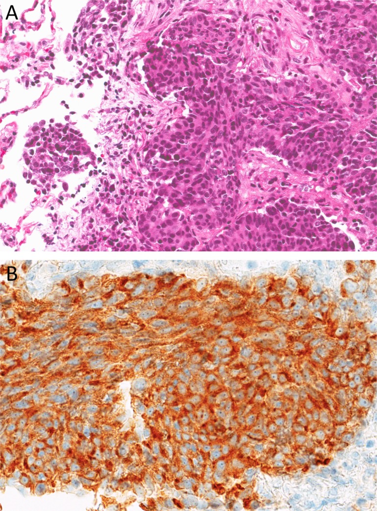
Figure 2.
Core biopsy findings. (A): Hematoxylin and eosin section shows consolidated lung parenchyma with invasive nests of neoplastic epithelioid cells within a surrounding desmoplastic stroma. The tumor cells have round‐to‐ovoid nuclei with dense chromatin, rare small nucleoli, and moderate amount of eosinophilic cytoplasm. Adjacent normal lung parenchyma is seen on the left (×200). (B): Immunohistochemical stain for synaptophysin demonstrates positive cytoplasmic granular staining (×400).

