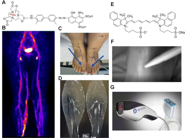
Figure 1.

Imaging procedures of 68Ga-NEB PET/MR/ICG fluorescence lymphangiography. Albumin-binding radiotracer 68Ga-NEB was injected subcutaneously into the bilateral first web spaces of the feet. Patients were requested to walk for 30 min and then PET image acquisition was performed. (A) Structure of 68Ga-NEB. (B) Representative maximum intensity projection of 68Ga-NEB PET with high signal intensity line of the lymphatic vessels acquired at 60 min after tracer administration. (C) Injection site of 68Ga-NEB to a patient with lymphedema. (D) A representative contrast enhanced 3D MR image of lymphatic vessels after subcutaneous injection of Gd-DTPA. (E) Structure of ICG. (F) Real time ICG lymphography acquired intraoperatively. (G) The fluorescence camera used for ICG imaging.
