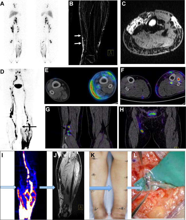
Figure 3.

Imaging guided surgery in a patient with stage II lower-limb lymphedema. (A) 99mTc-SC lymphoscintigraphy showed dermal backflow of the left lower-limb, asymmetrical distribution of bilateral lymph nodes and accumulation defect on the left side. (B–C) MR lymphangiography showed dilated and discontinuous lymphatic channels of the left medial calf (arrows). (D–H) 68Ga-NEB PET lymphangiography (60 min after tracer administration) represented “Honeycomb Pattern” with dilated dermal lymphatics, discontinuous lymphatic channels and compensatory vessels and lymph lakes of the left lower-limb (D), as well as different levels of dermal backflow (E and F) and lymph node accumulation defect on the affected left side (G and H). (I–L) A typical workflow of imaging guided surgery planning using preoperative 68Ga-NEB PET (I) and MRL (J) to discriminate the location of the lymphatic channels (K) for lymphatic venous anastomosis (L).
