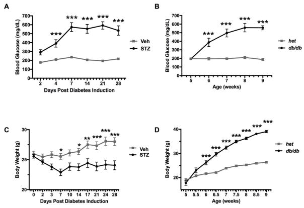
Figure 1.
Blood glucose (mg/dl) and body weight over time for diabetic and non-diabetic control mice. Panel A shows glucose levels over days following injection of A) Veh (n = 12) or STZ (n = 11). Panel B shows glucose levels in db/+ non-diabetic (n = 8) and db/db diabetic (n = 8) mice as a function of age. Body weights (mean ± SEM) are shown for the same mice injected with C) Veh (n = 12) or STZ (n = 11), and D) db/+ non-diabetic (n = 8) and db/db diabetic (n = 8) mice. Asterisks indicate significant difference between groups at each timepoint (***p < 0.001, ****p < 0.0001).

