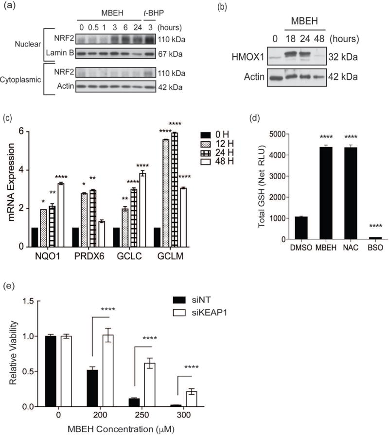
Figure 2. VIP exposure activates melanocyte NRF2 antioxidant responses, while constitutive NRF2 activation reduces MBEH-induced toxicity.

(a) Melanocytes were treated with MBEH (300 μM) for increasing periods up to 24 hours. Treatment with t-BHP (100 μM) for 3 hours served as positive control. Nuclear and cytoplasmic fractions were extracted and protein expression measured with Western blot analysis. Melanocytes were stimulated with MBEH (300 μM) for increasing periods up to 48 hours. (b) Total protein lysate was extracted for Western blot analysis of HMOX1 protein expression. (c) mRNA expression of NRF2-targets, NQO1, PRDX6, GCLC, and GCLM, was measured relative to housekeeping gene RPLP0 using quantitative RT-PCR. (d) Melanocytes were treated with MBEH (300 μM) for 20 hours and total GSH levels measured using a luminescence based assay. NAC (20 mM) and BSO (2 mM) treatment for 20 hours were used as positive and negative controls for GSH increase and depletion respectively. (e) Melanocytes were transfected with KEAP1 siRNA or NT siRNA for 48 hours followed by exposure to increasing concentrations of MBEH and relative viability measured after 24 hours. mRNA expression data and relative luminescence data are presented as mean ± SEM, n=3. The relative viability data is presented as mean ± SD. * p < 0.05; ** p < 0.01; *** p < 0.001; **** p < 0.0001. RLU, relative luminescence units; GSH, glutathione; NAC, N-acetyl cysteine; BSO, buthionine sulfoximine.
