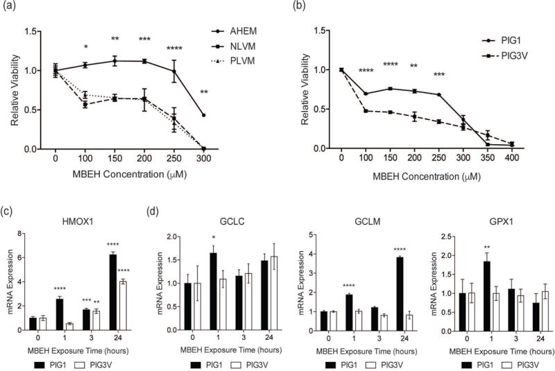
Figure 3. An impaired NRF2 response contributes to increased MBEH-induced toxicity in vitiligo melanocytes.

(a) Relative viability of normal adult human epidermal melanocytes (AHEM) and melanocytes derived from perilesional (PLVM) and non-lesional (NLVM) skin from a vitiligo patient were compared after exposure to increasing concentrations of MBEH for 96 hours. (b) Relative viability of immortalized control melanocytes (PIG1) and immortalized vitiligo melanocytes (PIG3V) compared after exposure to increasing concentrations of MBEH for 96 hours. PIG1 and PIG3V were stimulated with MBEH (300 μM) for increasing periods up to 24 hours and mRNA expression of (c) HMOX1 and (d) GSH pathway members GCLC, GCLM, and GPX1 was measured. The relative viability data is presented as mean ± SD. mRNA expression data are presented as mean ± SEM, n=3. * p < 0.05; ** p < 0.01; *** p < 0.001; **** p < 0.0001.
