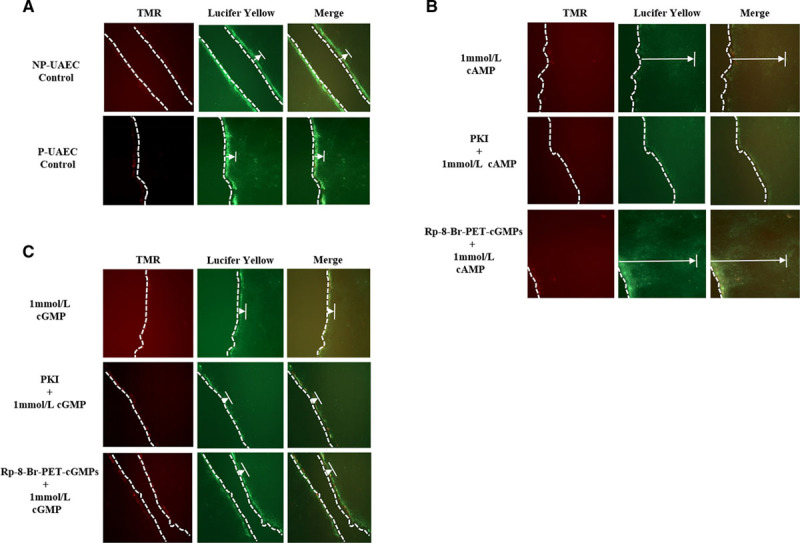
Figure 5.

Effects of PKA and PKG (protein kinase A and G) inhibitors on cyclic nucleotide–induced gap junction intercellular communication (GJIC). PKA, but not PKG-mediated processes, increased Cx43 (connexin 43) Lucifer yellow dye transfer. Representative fluorescence dye microscopy images: (A) untreated control nonpregnant and pregnant uterine artery endothelial cells (NP-UAECs and P-UAECs), (B) P-UAECs pretreated with 10 μmol/L of either PKA inhibitor (PKI; 1 hour) or PKG inhibitor (Rp-8-Br [8-Bromo]-PET-cGMPs; 1 hour) then 1 mmol/L 8-Br-cAMP for 30 minutes, and (C) P-UAECs pretreated with 10 μmol/L of either PKA inhibitor (PKI; 1 hour) or PKG inhibitor (Rp-8-Br-PET-cGMPs; 1 hour) then 1 mmol/L of 8-Br-cGMP for 30 minutes. Left, Illustrates tetramethylrhodamine (TMR) dextran staining at the cell membrane. TMR dextran is too large to pass through gap junctions (GJs) and, thus, serves as a measure of cell damage at the wounding site and a marker for Lucifer yellow entry. Middle, Illustrates Lucifer yellow dye passing through GJs into adjacent cells. Right, An overlapping merged photo of the TMR dextran and Lucifer yellow dye images. Dotted lines (—) denote the wound site, whereas the extended arrows (|←) denote the distance Lucifer Yellow dye traveled from the wound site.
