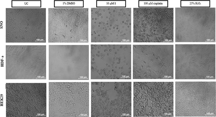
Fig. 3.
Light microscope images of malignant SNO, non-malignant HDF-a and HEK293 cells taken 24 h after treatment. All three cell lines were either untreated (UC) or treated with 1% DMSO, 10 µM complex 1, 100 µM cisplatin (apoptotic control) and 25% H2O2 (necrotic control). Images were taken at a magnification of ×200 on a Zeiss Axiovert 25 inverted microscope. Untreated and DMSO treated cells appear to be intact with no signs of cellular stress. Cells treated with complex 1 however show signs of cellular rounding and blebbing which resembles that of cisplatin. Necrotic cell death appears to be absent

