
Figure 2.
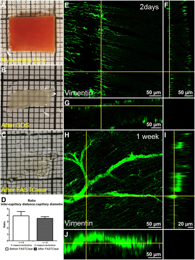
FASTClear provides an efficient myocardial tissue clearing protocol. (A–C) representative images of a myocardial slice before FASTClear, after SDS buffer treatment and after FASTClear protocol. (D) During tissue clearing the architecture and proportions of the myocardium is preserved and no changes in the inter-capillary distance to capillary diameter ratio were observed. Error bar = SEM. P > 0.05 T-test. (E–G) Orthogonal view of a myocardial slice after 48 hours antibody incubation, the labelling is limited to the surface of the preparation. (H–J) Orthogonal view of a myocardial slice after 1 week of antibody incubation. The labelling is homogeneously distributed and reached the core of the preparation. At least 3 slices were used for each experiment.