
Figure 3.
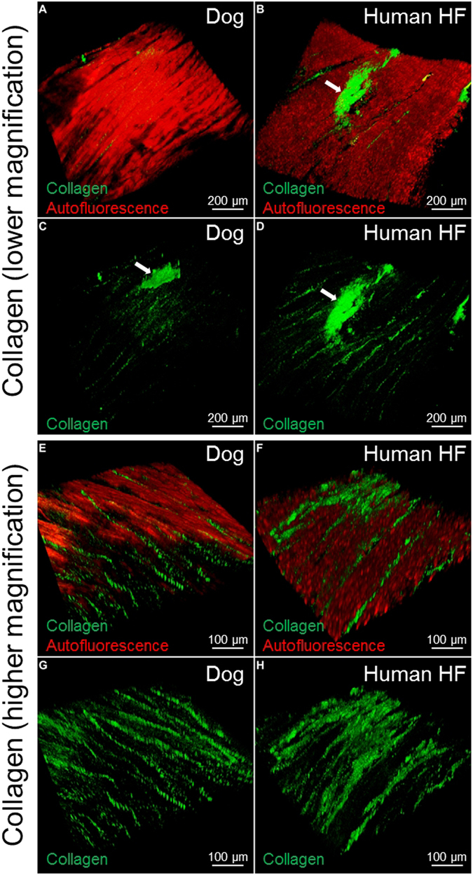
SHG imaging of Collagen type 1 in canine and human myocardial slices. Lower magnification of canine (A,C) and human (B,D) myocardial tissue. The autofluorescence (red) is used to show the structure of the myocardium; the collagen fibres (green) are located within the cardiac muscle fibres. The white arrows indicate the collagen in proximity to a large vessel. Higher magnification of healthy dog samples (E,G): the collagen is organised in strands between the muscle fibres; in human heart failure samples (F,H) the regular structure is partially lost and the collagen is less organised. At least 3 slices were used for each experiment.