
Figure 1.
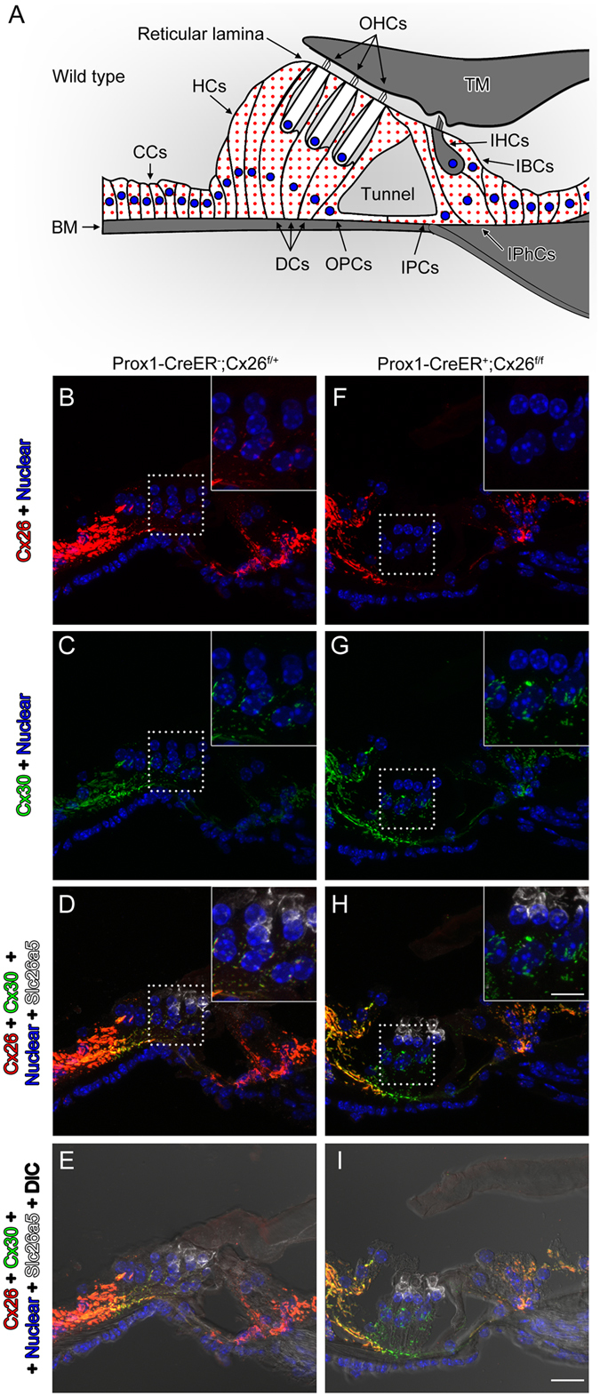
Specific deletion of Cx26 in Deiters’ and Pillar cells in Prox1-CreER; Cx26 f/f cKO cochleae. (A) Diagram of cross section of the organ of Corti. The cells containing red dots indicate Cx26 expressing cells in wildtype cochleae. Slc26a5 (prestin)-positive OHCs (by white) and the remaining regions (in grey) are indicated. DCs are located underneath of OHCs. IPCs and OPCs are located between IHCs and OHCs. (B–I) Cx26 (in red), Cx30 (in green), and slc26a5 (in white) expressions in middle turns of either wildtype (B–E) or Cx26 cKO cochleae (F–I) at P32 are shown. Counter-staining of nuclei is indicated in blue. Dashed boxes indicate the regions of the insets. Scale bar expresses 5 µm (in I) and 2.5 µm (in an inset in H). [See Fig. 2 below].