
Figure 2.
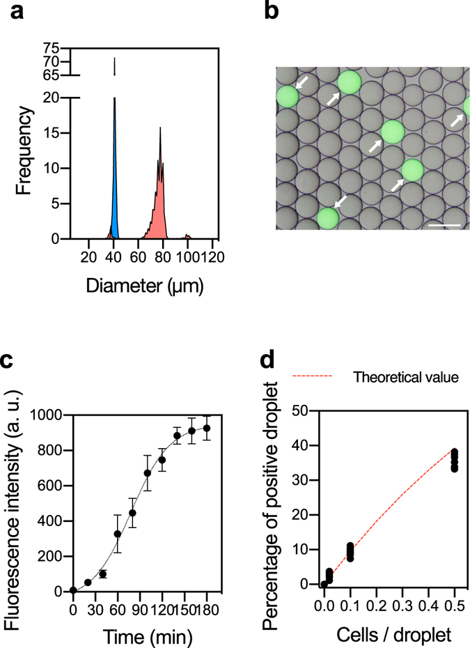
Droplet fusion and subsequent single-cell WGA in sd-MDA. (a) Histograms of droplets before (Cell lysate droplet: blue) and after fusion (SAG droplet: red). (b) Fluorescence image of droplets after the 1st-round MDA reaction. E. coli cells were introduced at 0.1 cells/droplet and their genomes were amplified for 2 h with Evagreen dye. Scale bar; 100 μm. (c) Time-dependent appearance of the fluorescence signal during amplification of single E. coli genome. All data are presented as averaged intensities of fluorescent positive droplets measured with SD, and 100 droplets were analyzed at each time point. (d) Relationship between introduced E. coli cell concentration and the number of fluorescent positive droplets.